Figure 2.
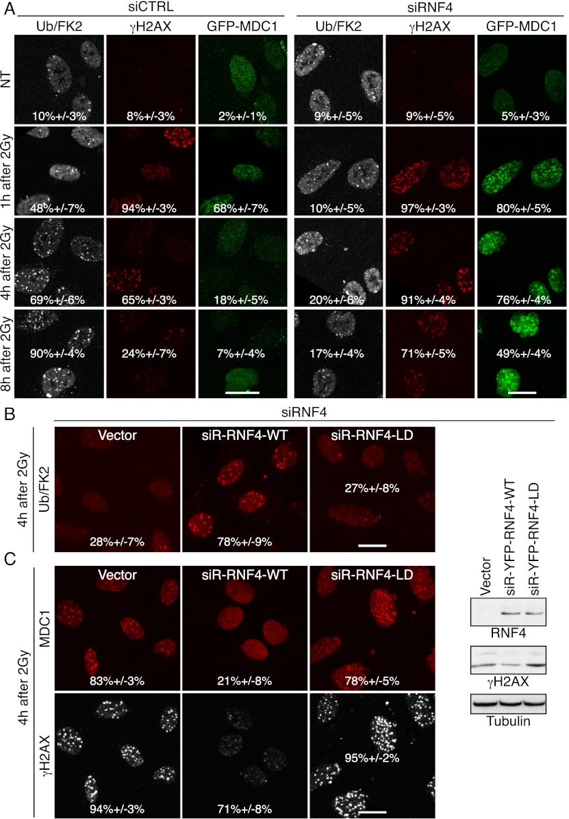
RNF4 depletion causes persistence of MDC1 and γH2AX foci. (A) U2OS cells stably expressing GFP-MDC1 were transfected with the indicated siRNAs, exposed to 2 Gy of IR, fixed after the indicated times, and analyzed by immunofluorescence. Quantification numbers represent proportions of cells showing Ub/FK2, γH2AX, or MDC1 foci ±SED (n > 100). (B,C) Cells stably expressing siRNA-resistant (siR) rat YFP-RNF4 wild type (WT) or LD or vector only were transfected with RNF4 siRNAs, exposed to 2 Gy of IR, fixed 4 h later, and processed as in A. MDC1 detection was with an anti-pSDTD-MDC1 antibody (Chapman and Jackson 2008) that detects constitutively casein kinase 2 phosphorylated MDC1. Quantifications for Ub/FK2 (B) and MDC1 and γH2AX (C) were done as in A. (Right panel) Corresponding samples were collected for immunoblotting. For additional time points, siRNA depletions, and resistant clones, see Supplemental Figures S2, A and B, and S11, A and B.

