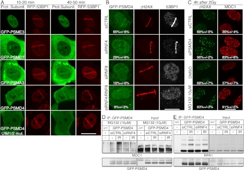
Figure 7.
Accumulation of the proteasome component PSMD4 at DNA damage sites requires its UIM domains, RNF4 and RNF8. (A) U2OS cells stably expressing RFP-53BP1 were transfected with GFP-PSME3, GFP-PSMD7, GFP-PSMA3, GFP-PSMD4, or GFP-PSMD4L218A,L220A,Y289A,M291A (UIM1/2 mut.). Forty-eight hours later, cells were laser-micro-irradiated, and live cells were imaged at the indicated times. (B) U2OS cells stably expressing GFP-PSMD4 transfected with siRNAs were laser-micro-irradiated, fixed after 2 h, and then analyzed by immunofluorescence. Quantifications: Numbers represent proportion of cells showing GFP-PSMD4 accumulation out of γH2AX-positive, ±SED (n > 100). (C) PSMD4 depletion or proteasome inhibition causes persistence of MDC1 and γH2AX IRIF. U2OS cells were transfected with siRNAs or treated with MG132 or DMSO immediately after irradiation (2 Gy of IR), fixed after 4 h, and analyzed by immunofluorescence as indicated. Quantifications: Numbers represent proportion of cells showing γH2AX or MDC1 foci, ±SED (n > 100). For time course and quantifications, see Supplemental Figure S10 (MDC1 detection was as in Fig. 2C). (D,E) PSMD4 interaction with MDC1 and RPA1 following IR is RNF4-dependent. (D) U2OS cells stably expressing GFP-PSMD4 were transfected with siRNAs and HA-tagged ubiquitin. Forty-eight hours later, cells were exposed to 10 Gy of IR and MG132. Extracts were prepared 4 h later and used for GFP-PSMD4 or GFP immunoprecipitations by GFP-Trap-A beads. Samples were analyzed by 4%–12% SDS-PAGE and immunoblotted as indicated. (E) As in D, but cells were not transfected with HA-Ub or treated with MG132. For siRNA depletions, see Supplemental Figure S11, A and C.

