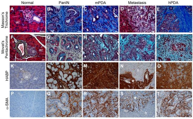
Figure 1. Evolution of the desmoplastic reaction in murine (mPDA) and human PDA (hPDA).
(A-E) Masson’s trichrome histochemistry shows robust collagen deposition (blue) at all stages of disease.
(F-J) Movat’s pentachrome histochemistry reveals collagen (yellow), GAGs and mucins (blue), and their co-localization (turquoise/green).
(K-O) Histochemistry with HA-binding protein (HABP) reveals intense HA content beginning with preinvasive disease (PanIN).
(P-T) Activated PSC express α-smooth muscle actin (α-SMA) and are abundant in preinvasive (Q), invasive (R) and metastatic mPDA (S) and hPDA (T), but not in normal pancreata (P). ac, acini; is, islet; d, duct; v, venule; h, hepatic parenchyma; *, metastatic lesions. Scale bars, 25 μm.
See also Figure S1.

