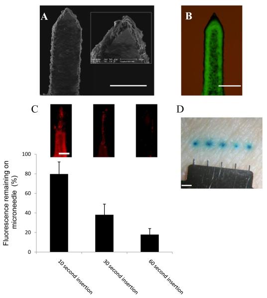
Figure 3. Coating microneedles and dissolution of coating in excised human skin.
(A) SEM images of a single MN coated with fluorescent nanospheres (bar = 200μm). (B) Bright field plus fluorescence microscopy of a single MN coated with fluorescent nanospheres (bar = 200μm). (C) Amount of fluorescently coated material remaining on MN surface following insertion into human skin; data presented as mean percentage fluorescence remaining ± SD (n=4), two replicates. Representative images of entire single needles from corresponding time points are shown as inserts (bar= 50 μm). (D) MNs coated with methylene blue demonstrate dissolution and deposition of the coated material within the skin (bar = 1mm).

