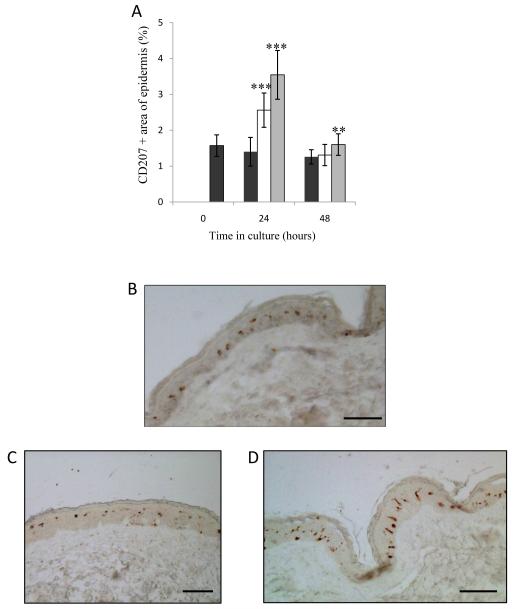
Figure 5. Area and distribution of LC in histological sections.
(A) The area of histological skin sections staining positive for CD207; Blank skin (black bars), H5 VLP MN treated (white bars), H1 VLP MN treated (grey bars). Data presented as mean ± SD (n=4), one replicate. Significance was determined relative to blank skin at corresponding time point (*p≤0.05 **p≤0.01, ***p≤0.001). (B-D) The distribution of LC in histological skin sections: blank skin (B); skin treated with H5 and H1 VLPs delivered from coated MNs (C & D respectively). All images taken from sections of cultured skin samples 24 hours post treatment (bar = 50μm in all cases).

