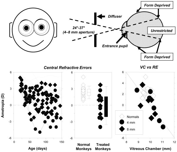
Figure 7.
Effects of peripheral form deprivation on refractive development.92 Top. Schematic illustrating the effects of the treatment lenses. Bottom left. Spherical-equivalent refractive corrections measured along the pupillary axis plotted as a function of age for the right eyes of individual control animals (thin gray lines) and treated monkeys (filled symbols) reared with diffusers with 4 mm (diamonds) and 8 mm apertures (circles). Bottom middle. Refractive errors for treated (diamonds, 4 mm apertures; circles, 8 mm apertures) and control animals at ages corresponding to the end of the period of peripheral form deprivation. The open and filled bars indicate the median and the 10th, 25th, 50th, and 90th percentiles for the control and treated monkeys, respectively. Bottom right. Vitreous chamber depth plotted as a function of spherical-equivalent refractive error for treated (diamonds, 4 mm apertures; circles, 8 mm apertures) and control animals at ages corresponding to the end of the treatment period. The line represents the results from the regression analysis of the data from the treated monkeys.

