Figure 2.
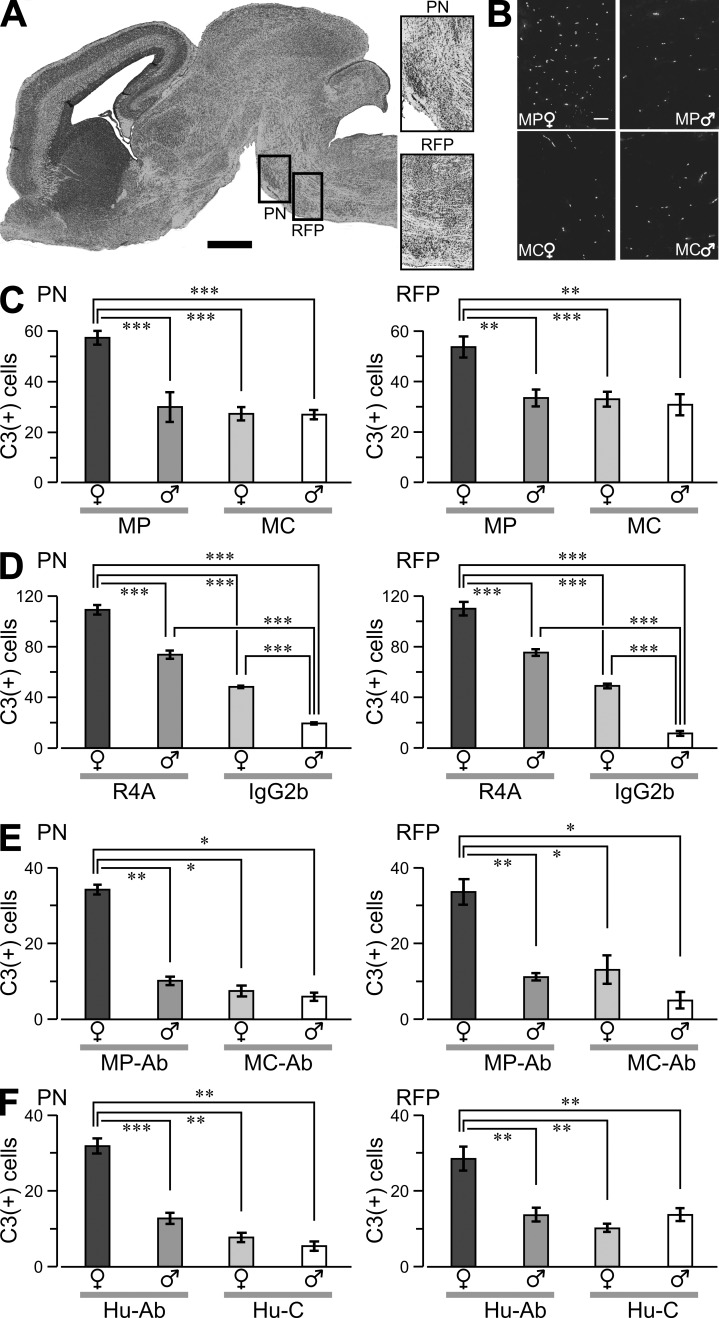
Neuronal death in fetal brains. (A, left) Sagittal whole-brain section (Nissl stained) of female fetus at E17. The boxes highlight the PN and RFP. (right) Representative magnified PN and RFP sections. (B) PN sections labeled for activated caspase-3 (C3+) in fetuses from MP- and MC-immunized dams. Bars: (A) 500 µm; (B) 50 µm. (C–F) Graphs show C3+ cells (mean ± SEM) counted in comparable regions (volume, 1.2 × 106 µm3) of the PN and RFP in the fetal brain. (C) Cell counts from MP and MC fetuses. Data are representative of three experiments. (D) Ex vivo brainstem slices from E17 fetuses were exposed to 100 µg/ml R4A or 100 µg/ml IgG2b for 12 h, fixed, and stained for C3+ reactivity. (E) Slices were treated with 200 µg/ml of purified serum DNARAbs (MP-Ab) or 200 µg/ml of mouse IgG (MC-Ab). (F) Slices were exposed to 400 µg/ml of human DNRAbs (Hu-Ab) or 400 µg/ml of human IgG (Hu-C). Sample sizes, 10–15 sections per column from three to four litters. *, P < 0.05; **, P < 0.01; ***, P < 0.001, ANOVA. Data are representative of two independent experiments.

