Figure 5.
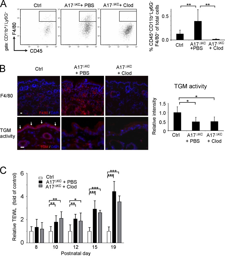
Infiltrating skin macrophages do not cause the skin barrier defects in A17ΔKC mice. For depletion of dermal macrophages, A17ΔKC mice were subcutaneously injected with clodronate-loaded liposomes (A17ΔKC + Clod) or PBS-loaded liposomes (A17ΔKC + PBS, Ctrl) into the back skin. (A) Flow cytometry of skin macrophages at P19 gated for CD11b+Ly6G− and further analyzed for skin macrophages (CD45+F4/80+; n = 5). (B) Immunofluorescence staining of A17ΔKC skin with anti-F4/80 antibodies to detect macrophages (top) and for TGM activity (bottom, arrows in Ctrl; n ≥ 5 per group). Bars, 20 µm. (C) TEWL from the back skin of A17ΔKC mice injected with clodronate-loaded (A17ΔKC + Clod) or control (A17ΔKC + PBS) liposomes and from control mice was detected from P8 to P19 (n ≥ 5 per group). Data are mean ± SD. *, P < 0.05; **, P < 0.01; ***, P < 0.001.

