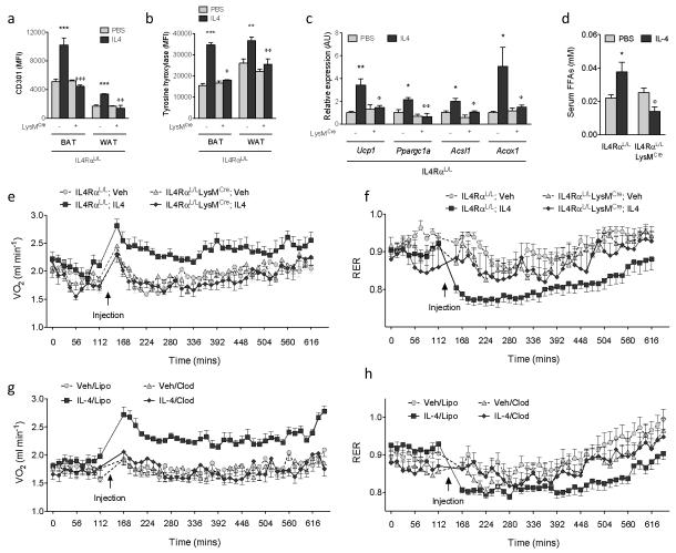
Figure 4. Alternative activation of macrophages increases energy expenditure.
a,b, Expression of alternative activation marker CD301 (a) and TH (b) in adipose tissue macrophages from IL4RαL/L and IL4RαL/LLysMCre mice treated with vehicle (Veh) or IL4 for 6 hours at 22 °C (n=4-5 per genotype and condition). c, Real-time PCR for thermogenic genes in BAT of IL4RαL/L and IL4RαL/LLysMCre mice treated with Veh or IL4 for 6 hours at 22 °C (n=4-5 per genotype and condition). d, Serum free fatty acid (FFA) levels in IL4RαL/L and IL4RαL/LLysMCre mice treated with Veh or IL4 for 6 hours at 22 °C (n=4-5 per genotype and condition). e, f, Quantification of energy expenditure in IL4RαL/L and IL4RαL/LLysMCre mice treated with vehicle (Veh) or IL4 (n=7-9 per genotype and condition). (e) Oxygen consumption (VO2) and (f) respiratory exchange ratio (RER). g, h, Quantification of energy expenditure in WT mice after macrophage depletion (n=8 per condition). Mice were injected with empty liposomes (Lipo) or clodronate-containing liposomes (Clod) 24 hours prior to energy expenditure studies. All data were collected during the light cycle. *P < 0.05, **P < 0.01, ***P < 0.001 compared to IL4RαL/L with Veh. φP < 0.05, φφP < 0.01, φφφP < 0.001 compared to IL4RαL/L with IL4.

