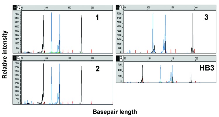
Figure A1.
Capillary electropherograms of Plasmodium microsatellite analysis. The x axis represents fragment size in bases and the y axis represents fluorescence intensity. The P. falciparum microsatellites products C13M30 and PFPK (black peaks) and TA81 and C4M8 (blue peaks) from amplification genomic DNA from patients 1, 2, one of the unrelated patient controls, and control P. falciparum clone HB3. Each microsatellite has ≥10 alleles. Patients 1 and 2 have identical microsatellites at all 4 loci (Table).

