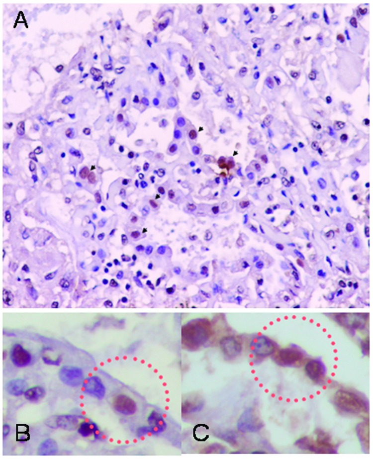
Figure 3.
Immunohistochemical analysis showing influenza A antigen-specific staining in nuclei of cells lining the alveoli (A). To identify the cell type, slides from consecutive sections were stained with anti-influenza A antibody (B) and double-stained with antiinfluenza A and antisurfactant antibodies (C). The sections were mapped, and the same area in each section was examined. Viral antigen-positive cells were stained both intranuclearly with antiinfluenza antibody and intracytoplasmically with antisurfactant antibody, indicating that the viral antigen-positive cells were type II pneumocytes. Viral antigen-positive cell are marked by circles (magnification x400).

