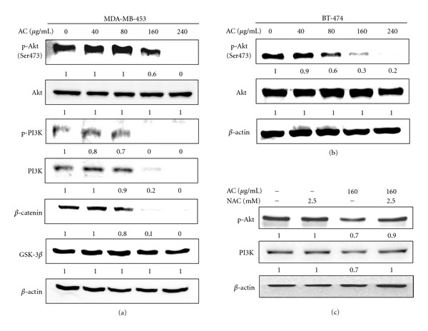
Figure 4.

AC treatment suppressed the phosphorylation of PI3K/Akt and GSK-3β/β-catenin in HER-2/neu-overexpressing breast cancer cell lines. (a) MDA-MB-453, (b) BT-474, and (c) 2.5 mM NAC pretreated MDA-MB-453 cells were treated with or without AC (40–240 μg/mL) for 24 h. The levels of phosphorylated PI3K (p-PI3K) and Akt (p-Akt, pSer 473 Akt) were evaluated using phosphorylated antibodies specific to PI3K and Akt in an immunoblot analysis. The total PI3K and Akt levels were assessed as the loading control. The levels of indicated proteins in the cell lysates were analyzed with specific antibodies, and the amount of β-actin was used as an internal control for sample loading. The photomicrographs shown in this figure are from one representative experiment that was performed in triplicate with similar results. Relative changes in protein bands were measured using densitometric analysis; the control was 1.0-fold, as shown immediately below the gel data. The results are presented as the mean ± SD of three assays. *Significant difference in comparison to the control group (P < 0.05).
