
Fig 3.
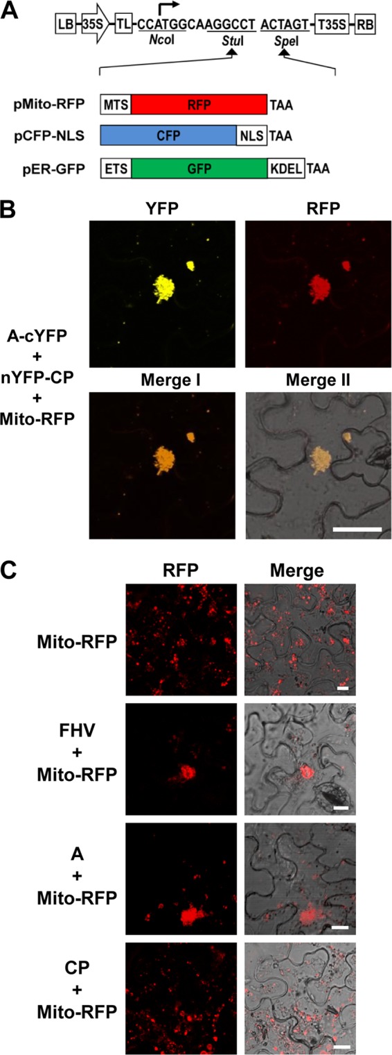
Protein A-CP interaction occurs on the mitochondria. (A) Schematic representation of binary vectors designed to express fluorescent subcellular markers. Three different subcellular marker genes were fused in-frame into the vector using StuI and SpeI sites. Plasmid pMito-RFP was designed specifically for mitochondria to emit red fluorescence by fusing ORF of red fluorescent protein (RFP) to mitochondrial targeting signal (MTS), plasmid pCFP-NLS was designed specifically for the nucleus to emit cyan fluorescence by fusing the ORF of cyan fluorescent protein (CFP) to the nuclear localization signal of simian virus 40 T antigen (NLS), and plasmid pER-GFP was specifically designed for the ER to emit green fluorescence by fusing the ORF of green fluorescent protein (GFP) to the ER targeting signal (ETS) and ER retention sequence (KDEL). (B) Visualization of subcellular localization of the protein A-CP interaction. A mixture of agro-constructs as indicated on the left side of the image was delivered into N. benthamiana leaves by agroinfiltration. The fluorescence signals were observed in the epidermal cells using confocal microscopy at 3 dpi. Bar, 30 μm. (C) Protein A mediates the clustering of mitochondria in plant cells. N. benthamiana leaves were infiltrated with the indicated agrotransformants. FHV denotes a mixture of F1 and F2 agrotransformants (3). The fluorescence signals were observed in the epidermal cells using confocal microscopy at 3 dpi. Mitochondria emit red fluorescence. Bar, 15 μm. Agrotransformants of protein A fused to YFP (A-YFP) and CP fused to GFP or RFP (GFP-CP or RFP-CP) were mixed with agrotransformants expressing the desired fluorescent subcellular markers and infiltrated into N. benthamiana leaves. The fluorescence signals were observed in the epidermal cells using confocal microscopy at 3 dpi. Mitochondria, nuclei, and ER emit red, cyan, and green fluorescence, respectively. Bar, 15 μm.