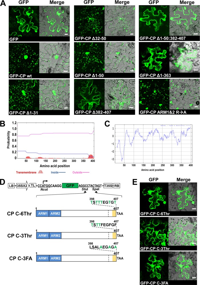
Fig 7.
Identification of the CP domain required for specific subcellular localization. (A) CP mutants were fused to GFP as shown in Fig. 5A to C. The indicated GFP-tagged CP mutants were expressed in N. benthamiana leaves by agroinfiltration. The GFP signals were observed in the epidermal cells using confocal microscopy at 3 dpi. Bar, 15 μm. (B and C) Prediction of the TMD (B) and the hydrophobic domain (C) of FHV CP. The TMHMM v.2 and ProtScale (the Kyte and Doolittle method) programs were used to predict the TMD and the hydrophobicity of CP, respectively. (D) Binary plasmids designed to express CP containing mutations (indicated in green font) in the C-terminal hydrophobic domain as GFP fusions. (E) After agroinfiltration of C-terminal mutants of CP, GFP signals were observed in the epidermal cells using confocal microscopy at 3 dpi. Bar, 15 μm.

