Fig 8.
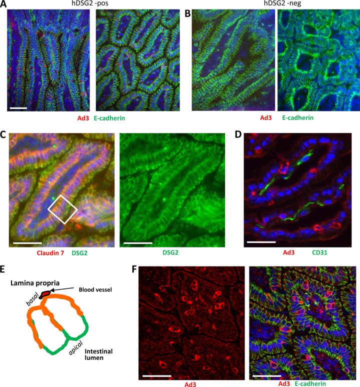
Uptake of Ad3-GFP by intestinal epithelial cells after intravenous injection. Ad3-GFP (1 × 10⁹ PFU/mouse) was injected into the tail vein of hDSG2-transgenic mice (hDSG2-pos) and nontransgenic littermates (hDSG2 neg). Two hours later, intestines were harvested and analyzed. (A and B) Intestine sections of hDSG2-positive (A) and hDSG2-negative (B) mice stained for Ad3 fiber knob (red) and E-cadherin. (C) Localization of hDSG2 (green) with regard to epithelial junctions. Junctions are marked by claudin 7 (red). Overlapping colors appear in orange. (D) Staining for Ad3-GFP (red) and blood vessels (marked by the endothelial cell marker CD31) (green). The scale bars are 20 μm. (E) Schematic representation of an intestinal villus (transverse section). Shown are two epithelial cells. The basal side faces the lamina propria, containing blood vessels. The apical side faces the intestinal lumen. hDSG2 is localized on all lateral membranes. Membrane areas that contain the adherens junction protein claudin 7 appear in orange. (F) Intestine section from hDSG2-positive mice injected with Ad3-GFP. Ad3-GFP appears in red; E-cadherin appears in green.

