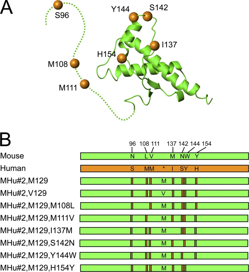
Fig 1.
(A) Structure of MHu#2(M129) based on that of mouse PrP (green; dashed line indicates unstructured region), with human PrP residues shown as orange spheres. (B) Schematic representation of mouse (green) and human (orange) PrP sequences, with human PrP residues (orange bars) highlighted in the chimeric sequences. Asterisk in human PrP sequence identifies the position of polymorphic residue 129 denoted methionine (M) or valine (V) in the chimeric sequences.

