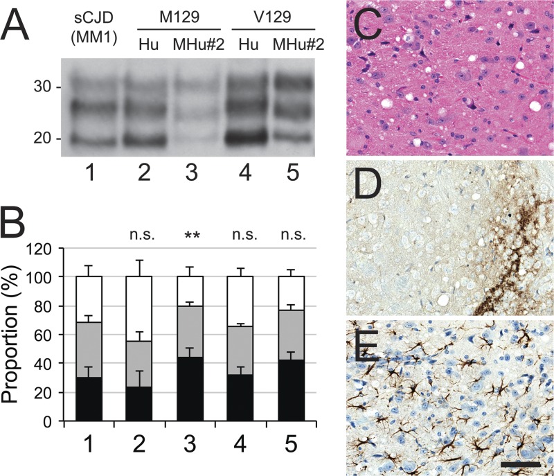
Fig 3.
Characterization after transmission of sCJD(MM1) prions to Tg mice expressing human (Hu) or chimeric (MHu#2) PrP with methionine (M) or valine (V) at polymorphic codon 129. (A) Immunoblot of brain homogenates: lane 1, inoculum; lane 2, Tg(HuPrP,M129)440; lane 3, Tg(MHu#2,M129)22372; lane 4, Tg(HuPrP,V129+/+)152; and lane 5, Tg(MHu#2,V129)6550 mice. Samples were treated with 100 μg/ml of PK for 1 h at 37°C prior to being loaded on gels and probed with the anti-PrP HuM-P antibody. Apparent molecular masses of migrated protein standards are shown in kilodaltons. (B) Proportions of unglycosylated (black), monoglycosylated (gray), and diglycosylated (white) PK-resistant PrP for each construct from multiple samples: 1, sCJD(MM1) (1 sample; 18 replicates); 2, HuPrP(M129) (5 samples; 8 total replicates); 3, MHu#2(M129) (6 samples; 11 total replicates); 4, HuPrP(V129) (1 sample; 4 replicates); 5, MHu#2(V129) (8 samples; 10 total replicates). Bars represent means, and error bars represent standard deviations; statistical difference from inoculum (lane 1) indicated above each bar: n.s., not significant; **, P < 0.01. (C to E) Micrographs of the hypothalamus of ill Tg Tg(MHu#2,V129)7110 mice stained with hematoxylin and eosin (H&E) (C) and stained immunohistochemically for PrPSc (D) or glial fibrillary acidic protein (E). Bar in panel E represents 100 μm and applies to all micrographs.

