Fig 3.

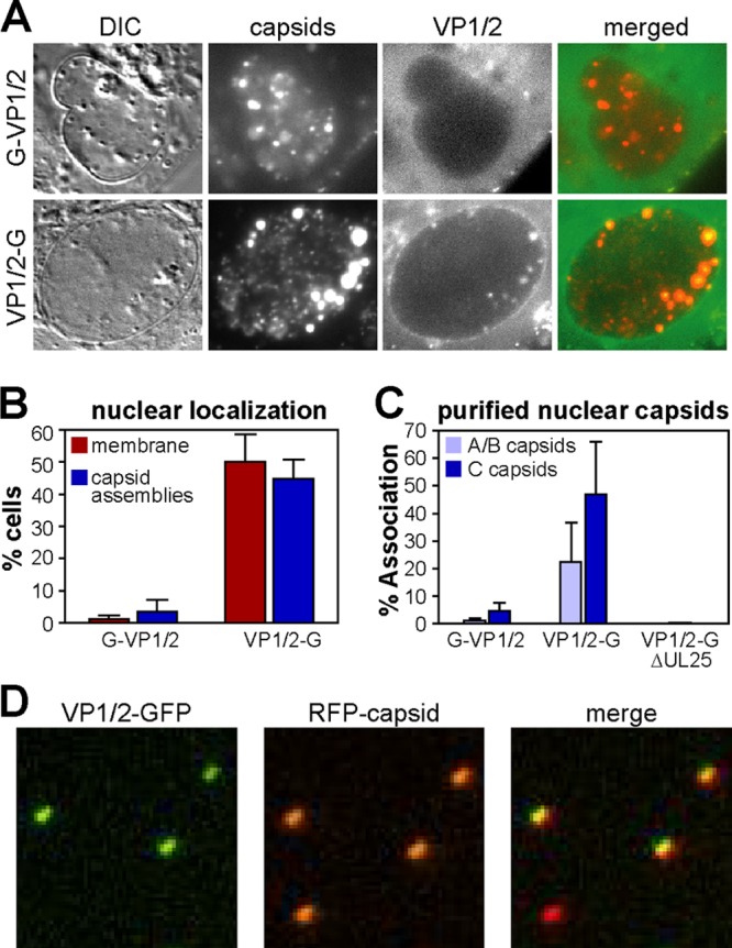
Nuclear localization of the VP1/2 carboxyl terminus. (A) Vero cells were infected with PRV-GS909 (G-VP1/2) or PRV-GS1903 (VP1/2-G). Images of infected Vero cell nuclei document the selective colocalization of VP1/2-G emissions with intranuclear capsid assemblies at 7 to 8 hpi. (B) Summary of GFP fluorescence localization at the nuclear membrane or with capsid assemblies in infected cells, as documented in panel A and in Fig. 2B (n = 202 for PRV-GS909; n = 332 for PRV-GS1903). The error bars represent the standard errors of the means. (C) Percent of RFP-capsids emitting GFP fluorescence following isolation of A/B and C capsids from the nuclei of infected Vero cells by rate zonal centrifugation. Only A/B capsid data are provided for the VP1/2-G + ΔUL25 sample, as C capsids were not recovered in the absence of the UL25 gene product. A minimum of 400 capsids were analyzed per sample. Each experiment was performed a minimum of three times. The error bars represent the standard errors of the means between experiments. (D) Subregion of an example image used to measure the frequency of VP1/2 association with isolated C capsids. Three of the four capsids shown were scored positive for GFP emissions, which in this example (PRV-GS1903) indicated the presence of the VP1/2 carboxyl terminus.
