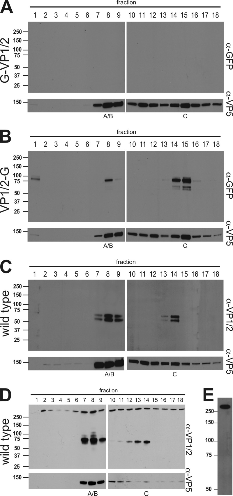
Fig 4.
Association of the VP1/2 carboxyl terminus with intranuclear capsids. Intranuclear capsids were harvested from infected Vero cells (A to C) or PK15 cells (D) and separated by rate zonal centrifugation. Fractions collected from the resulting sucrose gradient were precipitated with TCA and analyzed by Western blot. Fraction 1 represents the top of the gradient, and fraction 18 represents the bottom. Membranes were probed with antibodies directed against GFP, VP1/2, and the VP5 capsid protein. (A) PRV-GS909 (G-VP1/2). (B) PRV-GS1903 (VP1/2-G). (C and D) PRV-Becker (wild type). Peak fractions containing A/B or C capsids are indicated. (E) Extracellular PRV particles purified from supernatants of infected Vero cells were reacted with the VP1/2 antiserum. Molecular mass markers (kDa) are illustrated to the left of each panel.

