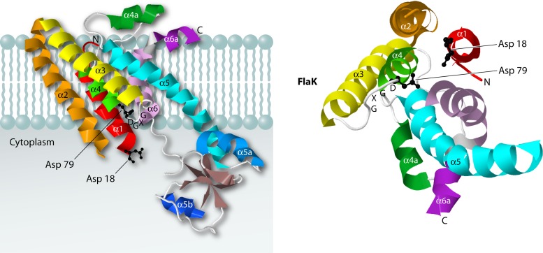
Fig 3.
Structure of the preflagellin signal peptidase FlaK of Methanococcus maripaludis. The structure on the left shows a side view of FlaK, and the structure on the right represents a view from the cytoplasmic side, with the cytoplasmic domain removed. Each of the six α helices is colored differently. The two catalytic Asp residues are shown as ball-and-stick models, in black. The crystal structure data were obtained using PDB accession number 3S0X, and JMol was used to generate the 3D structure images.

