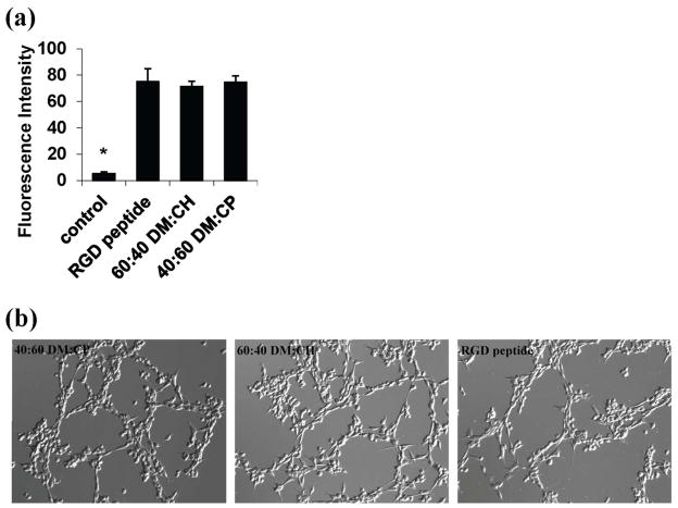
Figure 5.
Quantitative and qualitative evaluation of 3T3 fibroblasts cultured on surfaces modified with the best nylon-3 long polymers or with an RGD-containing peptide after a two-day incubation. (a) The vertical axis indicates fluorescence intensity based on live-cell staining; (*) indicates p < 0.0001 compared to RGD. (b) Bright field micrographs (100x total magnification) of 3T3 cells on the surfaces modified with the nylon-3 copolymers or the RGD-containing peptide.

