
Figure 2. Activity test of PknBSA-KD.
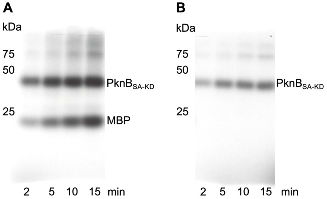
PknBSA-KD (25 ng) was incubated either with myelin basic protein (MBP; 1 µg) (A) or alone (B) together with γ33-ATP, MnCl2 and MgCl2 for the time indicated. Position and size (kDa) of molecular weight markers are indicated on the left side. Phosphorylation of MBP (A) and autophosphorylation (B) are visualized by autoradiography using direct-exposure film. The phosphorylation rate is increasing as a function of time in both experiments, demonstrating that the purified PknBSA-KD protein is active.