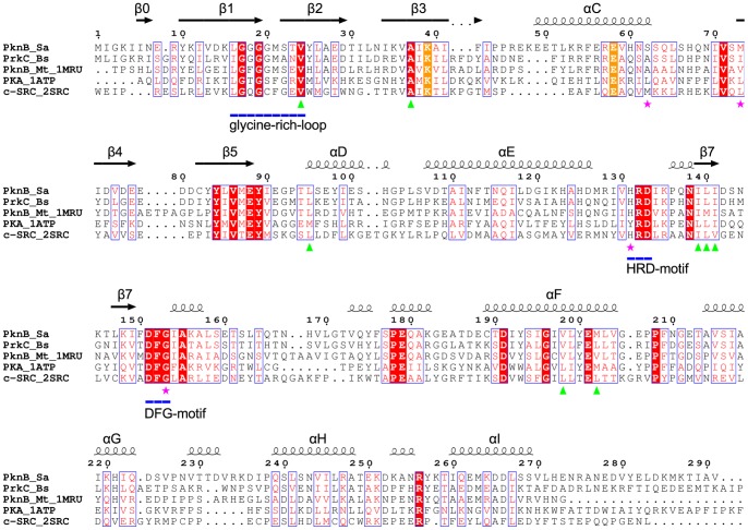
Figure 5. Structure-based sequence alignment.
Structure-based sequence alignment of the S. aureus PknB kinase domain with the kinase domains of B. subtilis PrkC (no structure available), M. tuberculosis PknB (PDB ID: 1MRU [24]), murine cAMP dependent Protein Kinase A (PDB ID: 1ATP [35]; PDB ID: 1CTP [34]) and human tyrosine protein kinase c-Src (PDB ID: 2SRC [36]). The secondary structure of PknBSA-KD is shown above the alignment and the numbering of the sequences corresponds to S. aureus as well. The HRD- and DFG-motifs and the glycine-rich loop are underlined in blue. The highly conserved residues Lys39 and Glu58 are marked in orange. Green triangles indicate the residues of the C-spine; magenta stars mark residues of the R-spine.

