
Figure 2. RPE fractions isolation scheme.
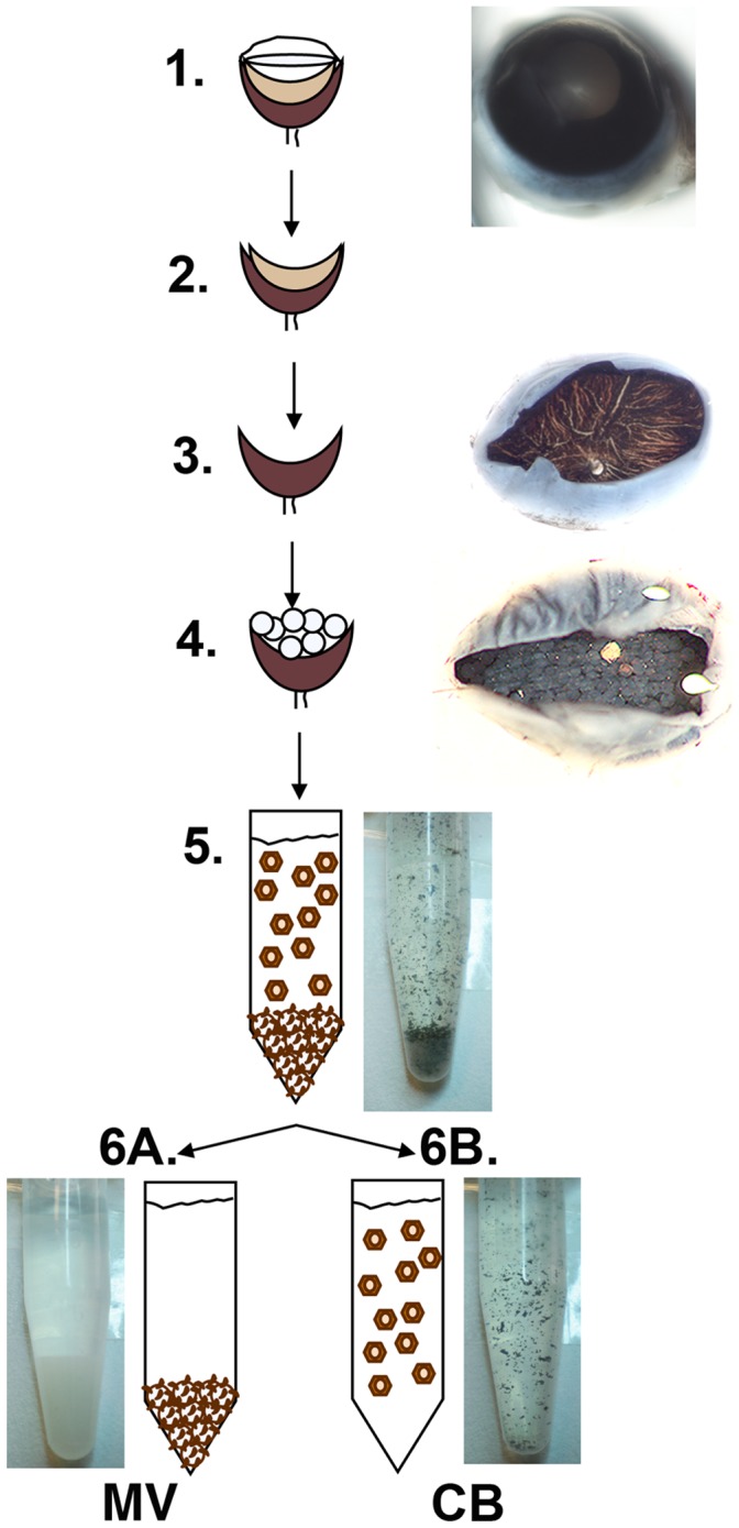
1-Mouse eyes are enucleated, 2-the anterior segments are surgically removed, and 3-the retina is removed after enzymatic treatment. 4- WGA-agarose beads are subsequently overlaid on the exposed RPE eyecups. After incubation, 5- the WGA-beads and RPE CB are gently scraped from the eyecups and collected into tubes. 6A- While WGA-agarose macrobeads are allowed to decant to the bottom of the tubes, 6B- the supernatant, with CB, is transferred to a fresh tube, and each fraction is washed and further processed for either biochemistry or morphology assays.