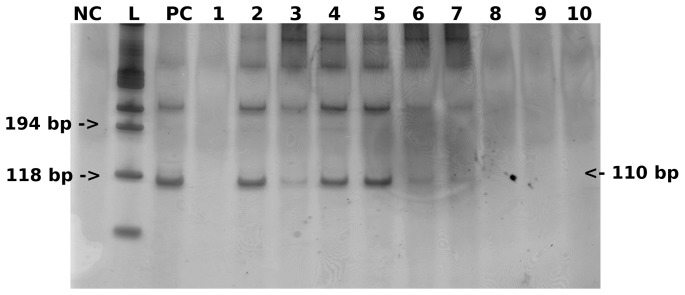
Figure 3. Visualization of 12 PCR assays with silver stained 8% polyacrylamide gel, showing the expected S. mansoni 110 bp DNA fragment in positive urine samples.
NC: negative control; L: 100 bp Ladder; PC: positive control; lines 2, 3, 4, 5, 6, and 7: S. mansoni positive urine samples; lines 1, 8, 9, and 10: S. mansoni negative urine samples.

