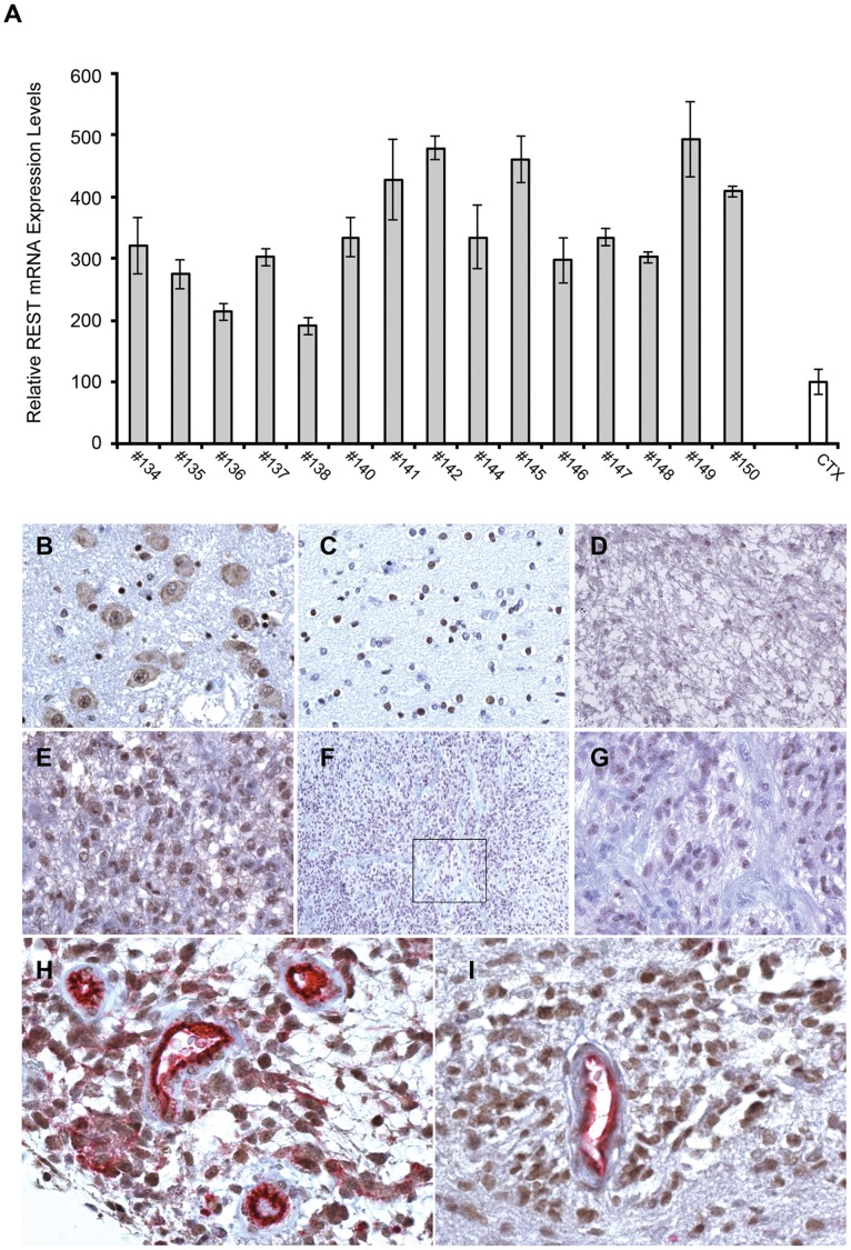
Figure 1. REST expression is elevated in human GBM.
(A) Quantitative RT-PCR analysis of REST mRNA level, relative to GAPDH, in human primary GBM specimens compared to pooled non pathologic human cerebral cortex tissues (CTX). Results are relative to three independent experiments. Data are means ± s.d. and were analyzed with Student’s t-test. (B-I) Representative images of immunohistochemical staining of REST in human non pathologic brain tissue and glioma specimens. (B) Neurons of pontine nuclei with nuclear and cytoplasmic staining. DAB, 400×. (C) Glial nuclei in the hemispheric white matter. DAB, 400×. (D) Diffuse astrocitoma: few nuclei are weakly positive. DAB, 200×. (E) GBM: all the nuclei in peculiar proliferating areas are intensely positive. DAB, 400×. (F) GBM: nuclei of hypercellular areas with high vessel density are intensely positive. DAB, 100×. (G) Inset of (f). DAB, 400×. (H and I) GBM: intense nuclear staining in perivascular cell cuffings for REST and Sox2 respectively (vascular tissue is revealed by CD-31 immunoreactivity). Double staining (REST-CD31, Sox2-CD31), 400×.

