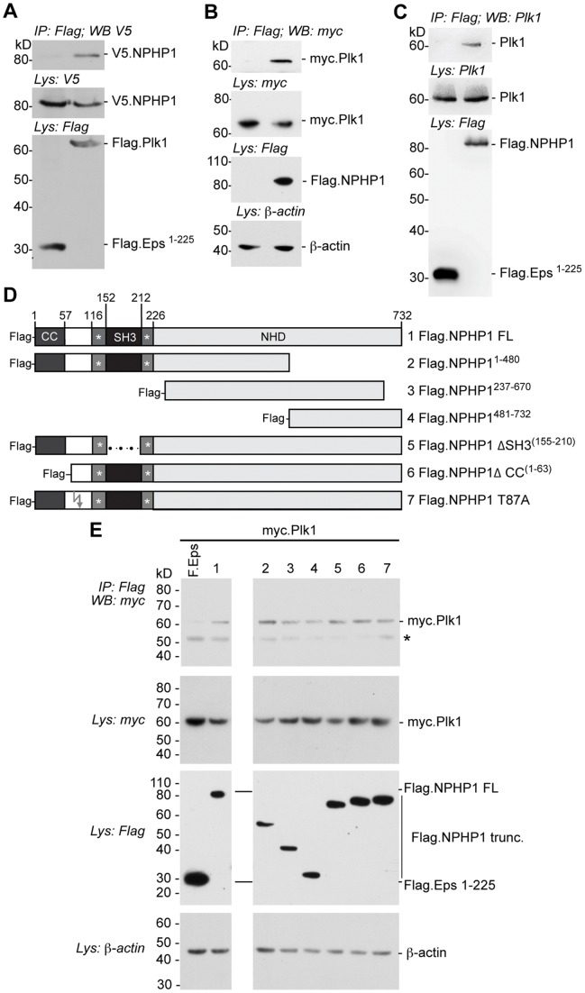
Figure 2. Plk1 associates with NPHP1.
A Western blot of immunoprecipitates (IP) or lysates (Lys) from HEK293T cells co-transfected with plasmids expressing V5-tagged NPHP1 and Flag-tagged Plk1 or negative control protein (Eps1–225 [13]). β-actin was assessed as a loading control. B Western blot of immunoprecipitates (IP) or cell lysates (Lys) from HEK293T cells co-transfected with plasmids expressing Myc-tagged Plk1 and Flag-tagged NPHP1 or empty Flag vector. C Western blot of immunoprecipitates (IP) or cell lysates (Lys) from HEK293T cells transfected with plasmid expressing Flag-tagged NPHP1 or the negative control protein (Eps1–225 [13]). Endogenous Plk1 was detected using a specific antibody against Plk1. D A panel of Flag-tagged NPHP1 derivatives, including truncations, internal deletions and a T87A mutant, was analyzed by co-immunoprecipitation with Myc-tagged Plk1. E Western analysis of immunoprecipitates (IP) or cell lysates (Lys) from HEK293T cells co-transfected with plasmids expressing Myc-tagged Plk1 and Flag-tagged NPHP1 constructs as indicated, or the Flag-tagged control protein (Eps1–225). * indicates immunoglobulin heavy chain.

