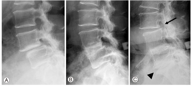
Fig. 4.
A 53-year-old woman with L4 spondylolisthesis. (A) A preoperative lateral radiograph shows a vertebral slip of 18% and intervertebral disc index of 0.13 at L4-5. (B) An 8-year postoperative radiograph shows complete union. The ceramic interspinous block is seen in the interlaminar space. (C) Adjacent disc degeneration is observed in a 16-year postoperative radiograph. The upper disc (L3-4) shows anterior vertebral slip of 16.7% (arrow). The lower disc (L5-S1) shows osteophyte formation and collapse (arrow head).

