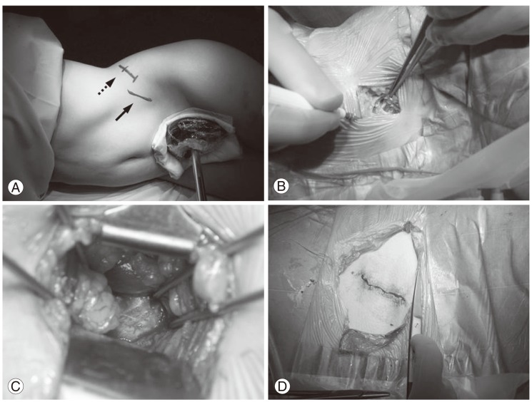
Fig. 1.
(A) A 4-cm skin incision (solid arrow) was made in the lateral abdominal region along the fibers of the external oblique muscle. The level of the L4-5 disc (dotted arrow) was located using the C-arm. (B) External oblique, internal oblique, and transverse abdominal muscles are dissected along the direction of their fibers. (C) The intervertebral disc is exposed using handheld retractors and Steinman pins. (D) Skin closure.

