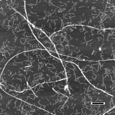
Figure 1.
Atomic-force microscopy of Aβ1–40 demonstrating assembly of amyloid fibrils from protofibrils. Preincubated Aβ1–40 (100 μM) was seeded with preformed fibrils amounting to ≈1% of the initial Aβ1–40 concentration. The aggregates were analyzed 7 days later. In this image, protofibrils appear as smaller and more flexible aggregates (≈4 nm in height) whereas the amyloid fibrils are substantially longer and more rigid (≈8 nm). (Bar = 200 nm.) [Reproduced with permission from ref. 8 (Copyright 1997).]

