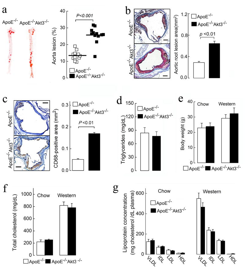
Figure 1. Deficiency of Akt3 promotes atherosclerosis in ApoE−/− mice.
(a) Representative images of en face oil red O staining of aortas of ApoE−/−Akt3−/− and ApoE−/− littermates (8 males and 4 females in each group), n=12. (b) (Left panel) Representative images of cross-sections of the aortic sinus stained with oil red O. (Right panel) Quantification of atheroma area. Six slides form different layers were taken for analysis from at least 3 hearts for each group. (c) (Left panel) CD68-positive macrophages (brown) in lesions of ApoE−/−Akt3−/− and ApoE−/− mice. (Right panel) Quantification of CD68-positive areas. (d) Fasting triglycerides levels of ApoE−/−Akt3−/− and ApoE−/− mice after 9 weeks on a Western diet. (e–g) Body weight, fasting cholesterol levels, and lipoprotein profiles from ApoE−/− and ApoE−/−Akt3−/− mice on a normal chow diet or a Western diet. Data represent means ± SEM.

