
Figure 2. Jejunal biopsies of normal (a) and diseased (b) intestines.
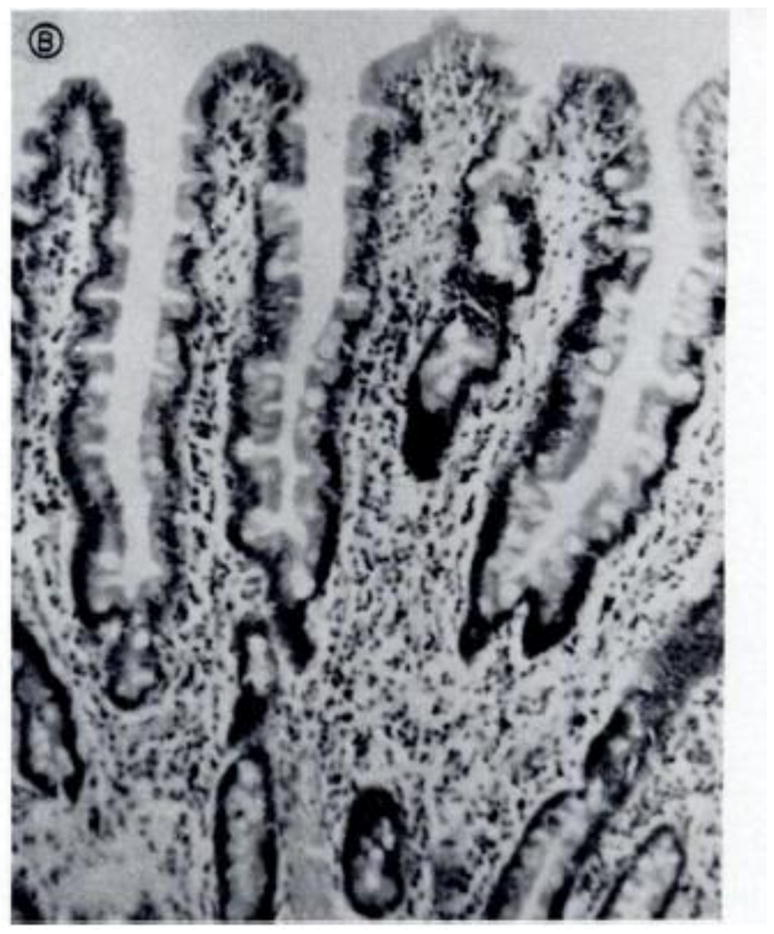

(a) Note the finger-like villi and short and narrow crypts of the normal intestine (a) compared with the severely flattened villi and inflammatory infiltration in the lamina propria and epithelium from the diseased tissue (b). This biopsy was taken from a Mexican adult with malnutrition, who may have had concurrent diarrhea. Reproduced with permission from [79].