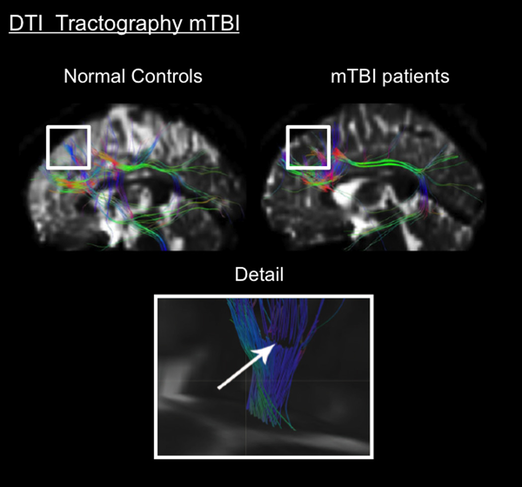
Figure 10.

Morphometric Imaging in Concussion. Top. DTI fiber tractography showing track fiber pattern to the left DLPFC in healthy controls and mTBI patients: 15 student-athletes (mean age 20.8 ± 1.7 years) who suffered from sport-related mTBI (collegiate rugby, ice hockey and soccer players). Statistical analysis demonstrated that these different patterns involved significant variations in diffusivity between these two groups; the mTBI group had decreased diffusivity. (From (Zhang et al., 2010a); permission pending).
Bottom. DTI fiber tractography detail of a mTBI patient at the level of the right semiovale center: Mild TBI patients possessed a GCS of 13–15 after a traffic accident, blow to the head or fall. Note the discontinuous characteristics of the fibers, hypothesized to be caused by trauma. (From (Rutgers et al., 2008); permission pending).
