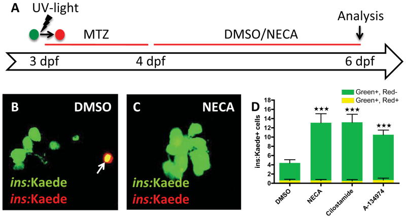
Figure 2. The hit-compounds increase regeneration, not survival, of β-cells.
(A) Schematic diagram for cell-labeling and assessment of β-cell survival/regeneration. To examine β-cell survival, we made use of the photo-convertible property of the fluorescent protein Kaede. At 3 dpf, before ablating the β-cells with MTZ from 3–4 dpf, we converted Tg(ins:Kaede)-expressing β-cells from green to red by exposing them to UV-light. After two days of regeneration (6 dpf), the surviving β-cells are red and green (yellow overlap), whereas the newly formed β-cells are green-only.
(B & C) Confocal images of DMSO- and NECA-treated larvae with Tg(ins:Kaede)-expressing β-cells at 6 dpf. Note that there is one β-cell that survived the ablation in this particular DMSO-treated larva (arrow in B), whereas there are no β-cells that survived the ablation in this NECA-treated larva (C).
(D) Quantification of β-cell regeneration (green bars) and β-cell survival (yellow bars) per larva at 6 dpf, following treatment with DMSO, NECA, Cilostamide, or A-134974 from 4–6 dpf. P < 0.0001; n = 10 larvae for each group. Error bars represent SEM. See also Figure S2.

