
Figure 2.
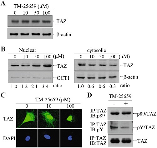
Nuclear localization of TAZ was enhanced by TM-25659. (A) C3H10T1/2 MSCs were treated with TM-25659 for 2 h. Whole cell extracts were resolved by SDS-PAGE and subsequently incubated with anti-TAZ antibody. (B) Nuclear and cytosolic proteins were prepared using the NE-PER kit (Pierce, Rockford, IL, USA) and 10 µg of nuclear protein and 50 µg of cytosolic proteins were used for SDS-PAGE and immunoblot analysis. OCT1 and β-actin were used as nuclear and cytosolic protein control respectively. (C) 3T3-L1 cells were treated with TM-25659 for 2 h and fixed for immunofluorescence assay using anti-TAZ antibody and Alexa 488-conjugated anti-rabbit IgG, followed by confocal microscopy. (D) 3T3-L1 cells were incubated with 100 µM TM-25659 for 2 h. TAZ protein was precipitated by incubation with anti-TAZ antibody and subjected to SDS-PAGE and immunoblotting with anti-pS89 TAZ antibody, phosphotyrosine antibody and anti-TAZ antibody.