
Figure 3.
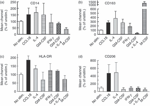
Effect of CCL18 on macrophage (MΦ) cell surface markers. Cultured monocytes (Day 2) were stimulated with 40 nm CCL18, 20 ng/ml interleukin-4 (IL-4), 20 ng/ml granulocyte–macrophage colony-stimulating factor (GM-CSF), alone or in combination, 20 ng/ml interferon-γ (IFN-γ), or 50 ng/ml macrophage colony-stimulating factor. On day 6 cells were harvested, stained with FITC-anti-CD14 and Cy5-anti-HLA-DR or FITC-anti-CD163 and allophycocyanin-anti-CD206 antibody and analysed by FACS. (a–d) Bars represent average values (– isotype control) ± SD of mean fluorescence intensities of three consecutive donors for CD14, CD163, HLA-DR and CD206 as indicated. Only CCL18-stimulated and IL-4-stimulated cells showed high expression of the mannose receptor CD206. Representative FACS histograms of these results are shown in the Supplementary material, Fig. S1.