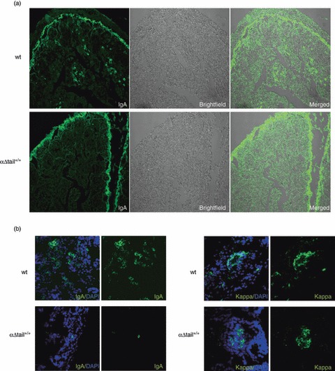
Figure 6.

Evaluation of the amount of IgA-secreting B cells (ASC) versus Igκ-secreting cells in mice small intestine. (a) Sections of small intestine villosities stained for IgA (left) or observed by light microscopy (middle) from wild-type (wt) or αΔtail+/+mice show IgA plasma cells only in wt tissues (as usual, the brush border is artefactually labelled with any fluorescent antibody) (original magnification × 10). (b) IgA versus Igκ labelling (Dapi counterstaining in blue) and representative profiles by confocal microscopy in wt and homozygous αΔtail+/+ tissues (original magnification × 63).
