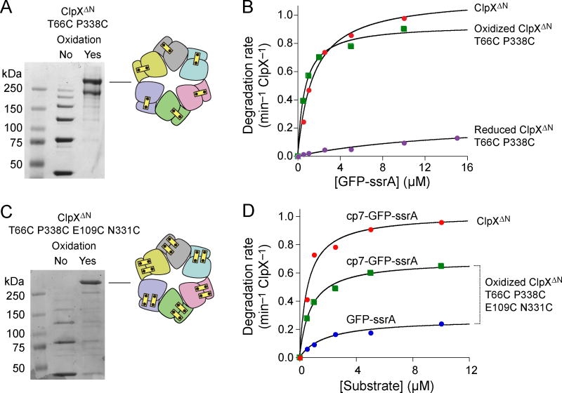
Figure 4.
Closed hexamers with disulfide bonds across all rigid-body subunit interfaces. (a) Non-reducing SDS-PAGE showing end-point disulfide-bond formation of T66C P388C ClpXΔN after oxidation with copper phenanthroline. (b) Michaelis-Menten plots of GFP-ssrA degradation by ClpXΔN, oxidized T66C P388C ClpXΔN, or reduced T66C P388C ClpXΔN (0.3 μM pseudo hexamer) and ClpP14 (0.9 μM). Symbols represent averages of three independent measurements. Table 1 lists values of KM and Vmax obtained by curve fitting. (c) Non-reducing SDS-PAGE showing end-point disulfide-bond formation of T66C P388C E109C N331C ClpXΔN after oxidation with copper phenanthroline. Symbols represent averages of three independent measurements. (d) Michaelis-Menten plots of GFP-ssrA or cp7-GFP-ssrA degradation by ClpXΔN or oxidized T66C P388C E109C N331C ClpXΔN (0.3 μM pseudo hexamer) and ClpP14 (0.9 μM). Table 1 lists KM and Vmax values.

