
Fig. 1.
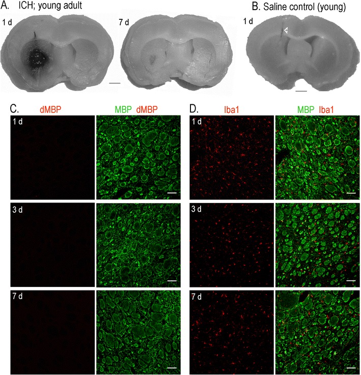
The collagenase model of ICH and the use of the contralateral striatum for animal- and age-matched comparisons. a A representative young adult rat showing that stereotaxic injection of type IV collagenase produced a hematoma that was restricted to the ipsilateral striatum. Much of the blood clot has been resorbed by day 7. b In a saline-injected control rat, the needle penetration track is seen (arrowhead) without bleeding in the striatum. c Representative confocal images of the contralateral striatum at 1, 3, and 7 days after ICH show normal white matter bundles labeled with myelin basic protein (MBP, green) and lack of degraded MBP (red). d In the contralateral striatum at 1, 3, and 7 days after ICH, microglia, identified with Iba1 (red) are of ramified morphology and broadly distributed between the white matter tracts. Scale bars, 500 μm