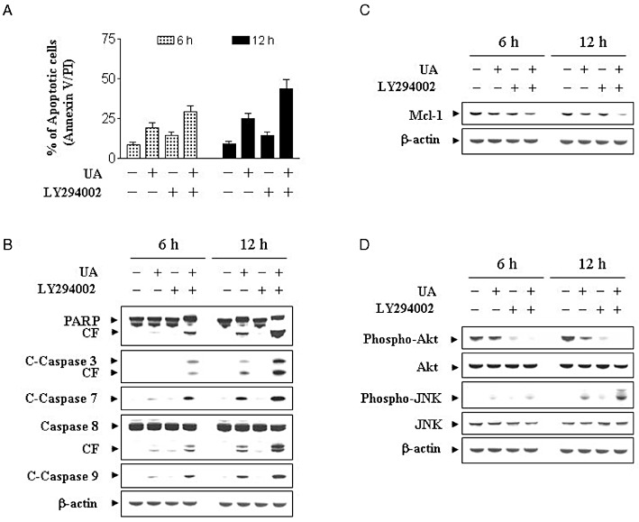
Figure 5.

Effects of the pharmacological inhibitor of PI3K/PKB on apoptosis induced by UA in U937 cells. U937 cells were pretreated with 20 µM of LY for 1 h followed by the addition of 10 µM of UA for 12 h. (A) Cells were stained with Annexin V/PI, and apoptosis was determined using flow cytometry as described in Methods. The values obtained from Annexin V/PI assays represent the means ± SD for three separate experiments. **Values for cells treated with UA and LY in combination were significantly greater than those for cells treated with UA alone by Student's t-test; P < 0.01. (B) Total cellular extracts were prepared as described in Methods and subjected to Western blot analysis using antibodies against PARP, C-Caspase-3, C-Caspase-7, caspase-8 and C-Caspase-9. Total cellular extracts were also prepared and subjected to Western blot assays using antibodies against Mcl-1 (C), and cell signalling proteins including phospho-PKB (Ser473), PKB, phospho-JNK and JNK (D). For the Western blot assay, each lane was loaded with 30 µg of protein; blots were subsequently stripped and re-probed with antibody against β-actin to ensure equivalent loading. Two additional studies yielded equivalent results.
