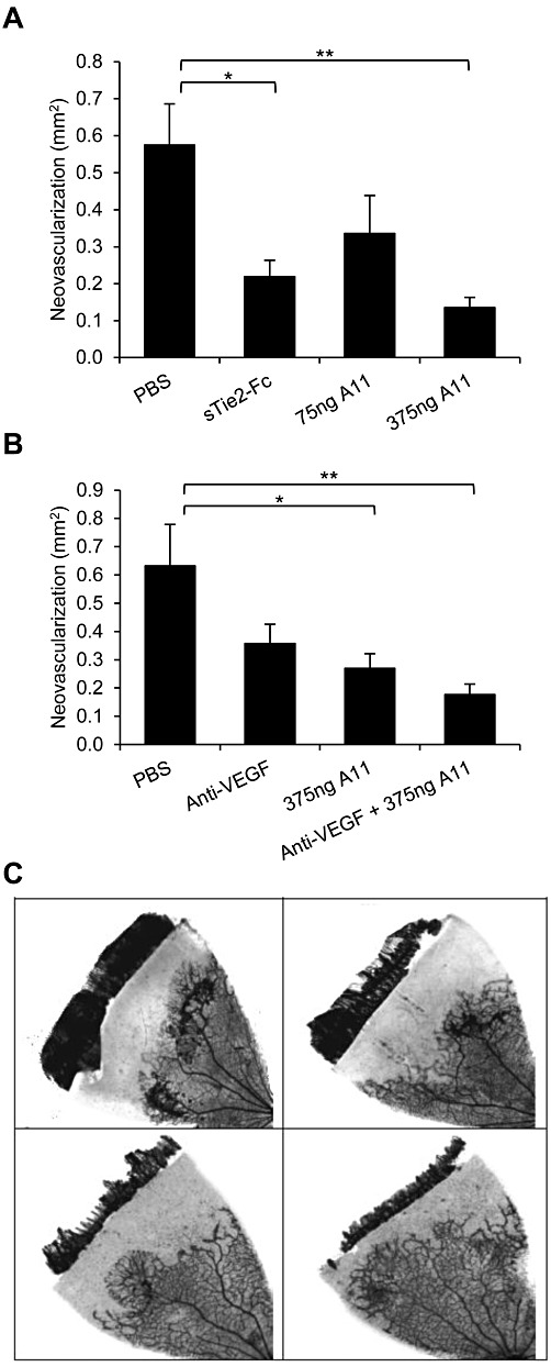
Figure 7.

Effect of peptide A11 on retinal neovascular growth. Retinal neovascularization was induced and assessed in Sprague–Dawley rats, using the OIR model. Data represent areas of abnormal vascular growth and are presented as mean ± SEM. (A) Rats received intravitreal injections of vehicle (PBS), peptide A11 at 75 or 375 ng per eye, or sTie2/Fc at 500 ng per eye as positive control. Both A11 at the higher dose and sTie-2/Fc yielded inhibition relative to PBS-injected eyes; **P < 0.01, *P < 0.05, significantly different as indicated; one-way ANOVA followed by Dunnett's post hoc test. (B). Rats received intravitreal injections of vehicle (PBS), anti-VEGF 500 ng per eye, peptide A11 at 375 ng per eye, or a combination of anti-VEGF and peptide A11 at 500 ng per eye and 375 ng per eye, respectively. Both peptide A11 and the combination of peptide A11 and anti-VEGF yielded inhibition relative to PBS-injected eyes; *P < 0.05, **P < 0.01, significantly different as indicated; one-way ANOVA followed by Dunnett's post hoc test. (C) Representative images of the degree of retinal neovascularization in the OIR model, following intravitreal injection of PBS (upper left), anti-VEGF (upper right), A11 (lower left) and A11 plus anti-VEGF (lower right).
