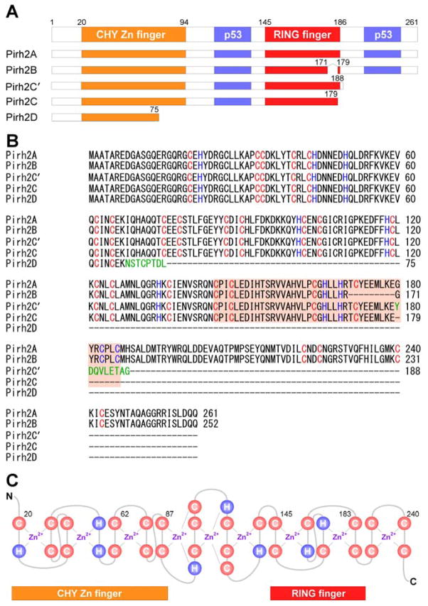
Fig. 1.
Schematic representation of Pirh2 and isoforms. (A) The domains of the human Pirh2 protein and the binding sites of p53 are indicated. (B) Sequence alignment of full-length Pirh2 and its isoforms using ClustalW2 multiple sequence alignment program. Pirh2C′ and Pirh2D have an additional unique amino acid (shown in green). (C) Secondary sequence organization of the CHY-zinc-finger/RING-finger domain. The cysteine and histidine are labeled as C and H, respectively. There are nine potential interleaved zinc binding sites.

