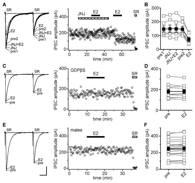
Figure 4. E2-induced IPSC suppression requires postsynaptic mGluR 1 and is sex-specific.
(A) Recording of E2-sensitive IPSCs in which E2 was applied in the presence and then absence of JNJ 16259685 (mGluR1 antagonist, JNJ, 0.2 μM); individual traces and time course are shown. Each point is an individual sweep and SR 95531 (SR, 2 μM) applied at the end of the experiment blocked IPSCs (also in C, E). JNJ blocked E2-induced IPSC suppression.
(B) Group amplitude data for all experiments as in A (n=6). Connected open symbols are individual cells; filled symbols are mean ± SEM for all cells (also in D, F).
(C) Recording of IPSCs with GDPβS in the recording pipette showing individual traces and time course. E2 had no effect on IPSCs with postsynaptic G protein signaling blocked by GDPβS.
(D) Group IPSC amplitude data for all experimetns as in C (n=10).
(E) Recording of IPSCs in a male tested with 100 nM E2 showing individual traces and time course. E2 did not affect IPSCs. Scale indicates 25 msec, 50 pA (also in A, C).
(F) Group IPSC amplitude data for all but one recording in males. E2 (10 nM, n=7 or 100 nM, n=15) had no effect on IPSC amplitude in 22 of 23 cells. In one recording not represented here, E2 produced a rapidly reversible decrease in IPSCs amplitude.

