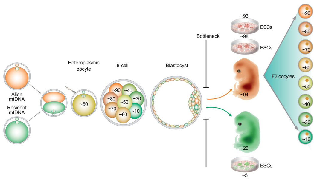
Figure 1. Schematic model demonstrating mtDNA segregation and bottleneck in primates.
Heteroplasmic rhesus monkey oocytes with equal mixture of two wild-type mtDNA haplotypes were constructed and mtDNA transmission to preimplantation embryos, fetuses and germ cells was followed. We demonstrate rapid segregation of mtDNA variants between daughter blastomeres in preimplantation embryos. However, fetuses and ESCs derived from these embryos shifted towards homoplasmic conditions. This bottleneck suggests that possibly a few cells within an ICM contribute to the somatic cell lineage of embryo proper. In contrast, individual fetal oocytes (F2) showed a wide range of heteroplasmy. This model also implies that the majority of ICM cells may contribute to the germline.

