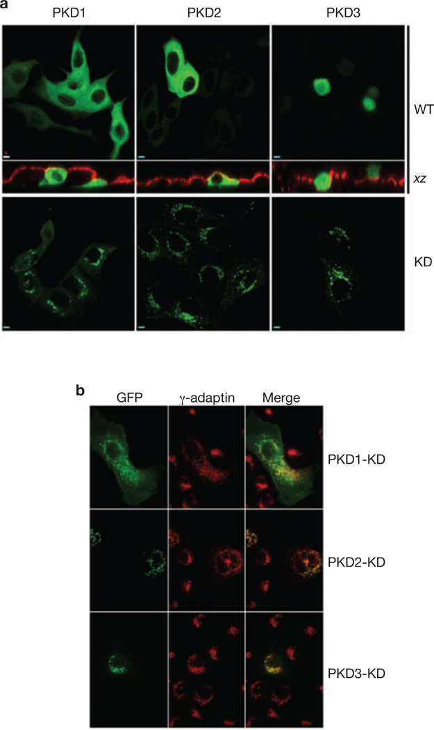
Figure 2.
KD isoforms of PKD localize to the TGN of MDCK cells. (a) MDCK cells were transiently transfected with WT or KD PKD1, PKD2 and PKD3, and cultured on Transwell filters (upper panels) or coverslips (lower panels). Cells expressing wtPKDs were co-stained with antibodies against gp135 to define the borders of the apical plasma membrane. Note that PKD1-WT and PKD2-WT were diffusely distributed throughout the cells, whereas PKD3-WT was present in the cytoplasm and the nucleus. The KD forms of these proteins were highly enriched on a perinuclear compartment. Scale bar, 5 µm. (b) Sub-confluent MDCK cells transiently expressing PKD1-KD, PKD2-KD or PKD3-KD were co-stained with antibodies against γ-adaptin, a TGN marker. Note that each kinase co-localized with a subset of membranes that were positive for γ-adaptin. Scale bar, 5 µm.

