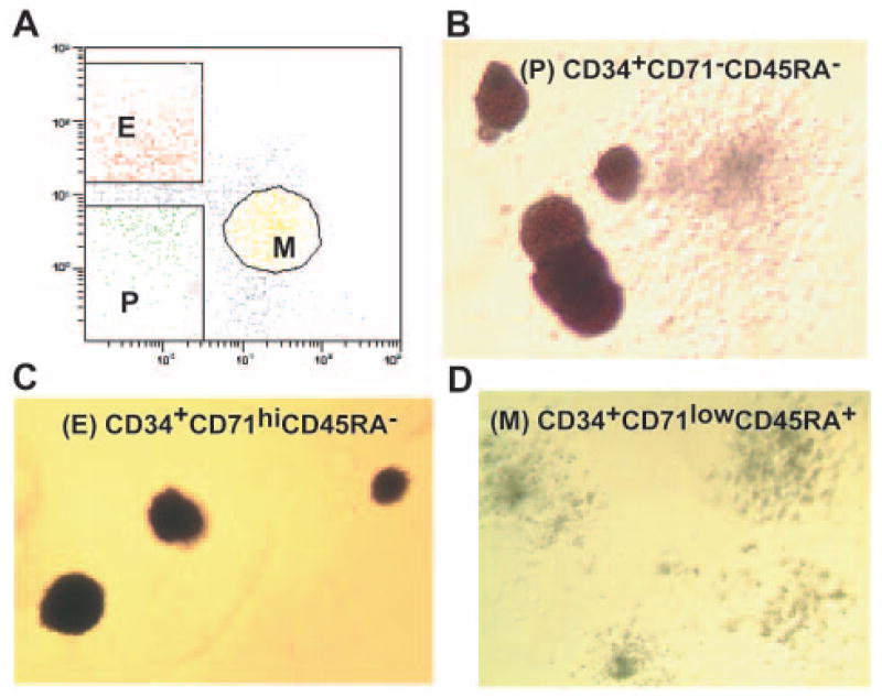
Figure 1.
Fluorescence-activated cell sorting separation and methyl-cellulose colony assays of bone marrow multipotential, erythroid and myeloid progenitors. (A): Three bone marrow (BM) populations isolated by flow cytometry: P (CD34+CD71−CD45RA− cells), E (CD34+CD71hiCD45RA− cells), and M (CD34+CD71lowCD45RA+ cells). The methylcellulose colony assays demonstrate that three populations of BM progenitors were highly enriched in primitive erythroid burst-forming units (BFU-E) and granulocyte-macrophage colony-forming units (CFU-GM) colonies from multipotential progenitors (average enrichment, 28 of 1,000 and 50 of 1,000 colonies, respectively) (B), more mature BFU-E and erythroid colony-forming units (CFU) colonies from erythroid progenitors (average enrichment for erythroid colonies, 160 of 1,000) (C), and granulocyte CFU, monocyte CFU, and granulocyte-macrophage CFU colonies from myeloid progenitors (average enrichment for myeloid colonies, 45 of 1,000) (D). Abbreviations: E, erythroid progenitors; M, myeloid progenitors; P, multipotential progenitors.

