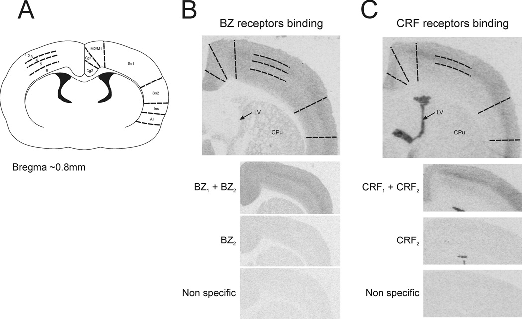
Figure 2. Cortical BZ and CRF receptors binding summary.
A) Scheme of the rat brain coronal section showing the cortical areas where BZ and CRF receptors were measured. B) Representative pictures for BZ receptors binding; total binding (BZ1+BZ2), binding after zolpidem displacement (BZ2) and binding after clonazepam displacement (non specific). C) Representative pictures for CRF receptors binding; total binding (CRF1+CRF2), binding after antalarmin displacement (CRF2) and binding after astressin displacement (non specific). Cg1 and Cg2, cingulate cortex area 1 and 2; M2/M1, primary and secondary motor cortex; Ss1 and Ss2, somatosensory cortex 1 and 2; Ins, insular cortex; AI, agranular insular cortex; 1–6, cortical layers 1 to 6; LV, lateral ventricle; CPu, striatum.

