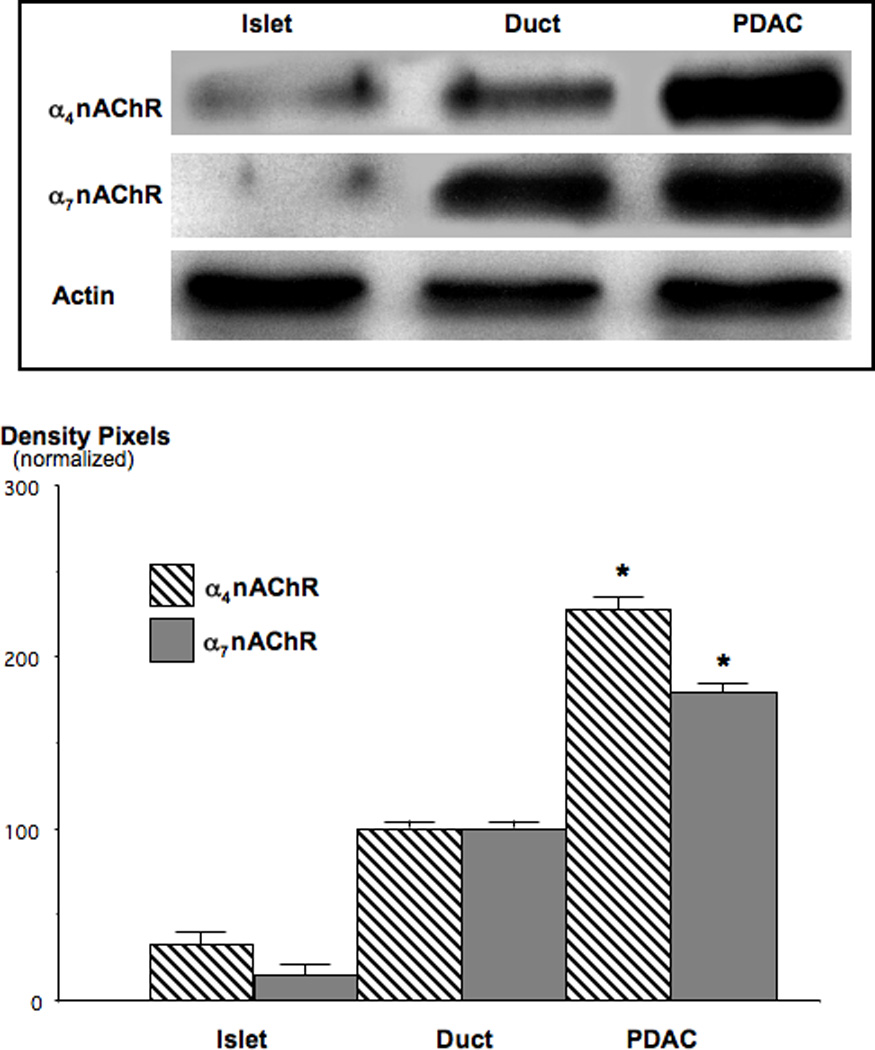
Figure 6.

Western blots, showing the upregulation of α4-and α7 nAChR subunit proteins in NNK-induced PDAC. Expression levels of the α4nAChR was increased 2.3-fold (*: p<0.001) and that of the α7nAChR 1.8-fold (*: p<0.001) over levels observed in duct epithelial cells. Data in the graph are mean values and standard errors of five densitometric readings per band of ratios of nAChR protein over actin and normalized with levels in duct epithelial cells set as 100%. Each Western blot was conducted three times with similar data.
