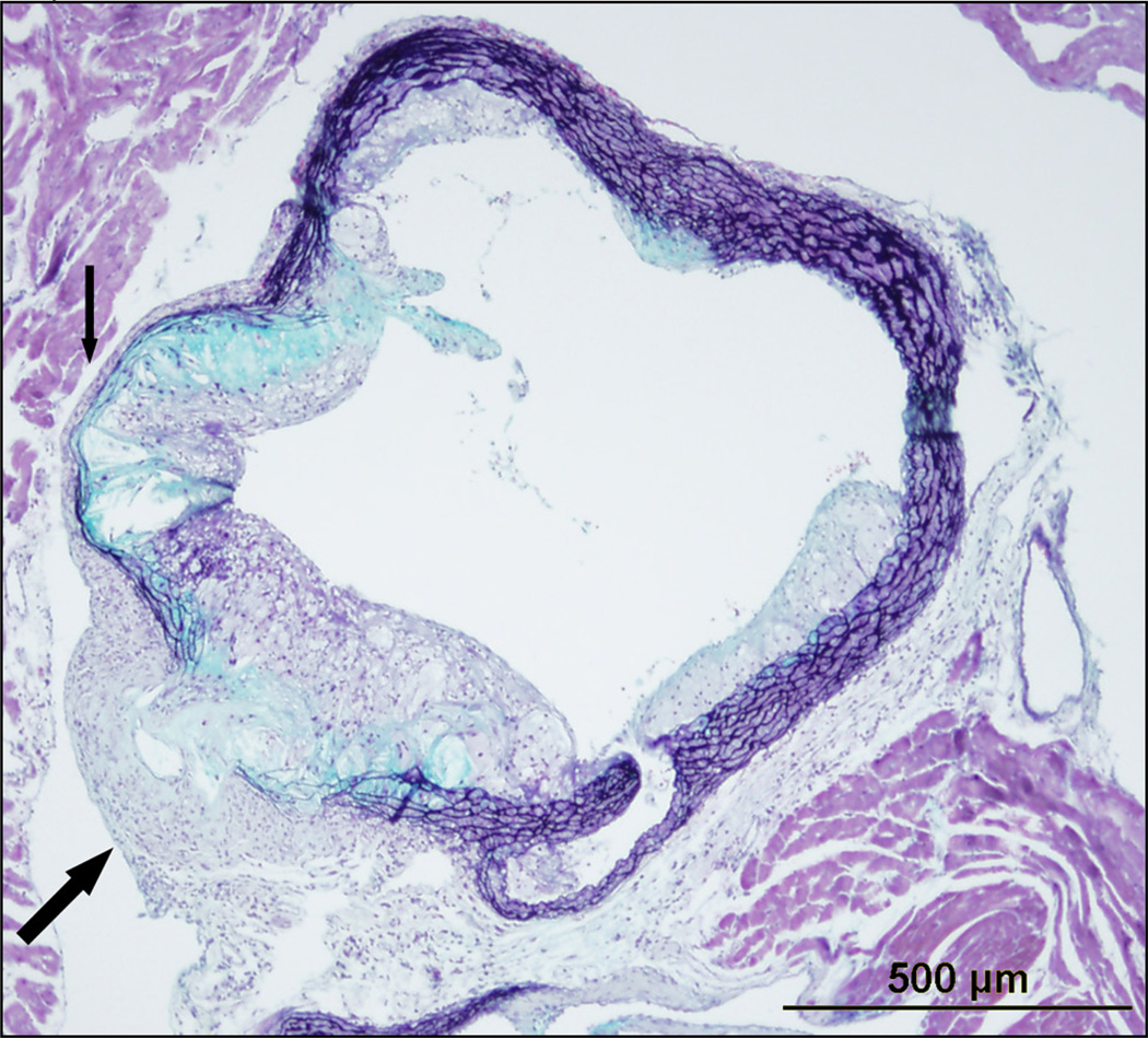
Figure 1. Movat’s stain of a cross section of a 24 week old Ldlr−/− mouse fed a Western diet for 16 weeks.
As indicated by the small arrow, there is thinning of the media and breakdown of the internal and external elastic lamina in the setting of advanced atherosclerotic plaque. The large arrow points to an advanced atherosclerotic lesion that has breached the internal elastic lamina, media, and external elastic lamina with evidence of necrotic core and cholesterol crystals within the adventitia. This breach enables emigration of intimal macrophages, dendritic cells, and lymphocytes to the adventitia and compromises the barrier status of the media. There is compensatory thickening of the adventitia in this region which likely serves to contain the breached media.

