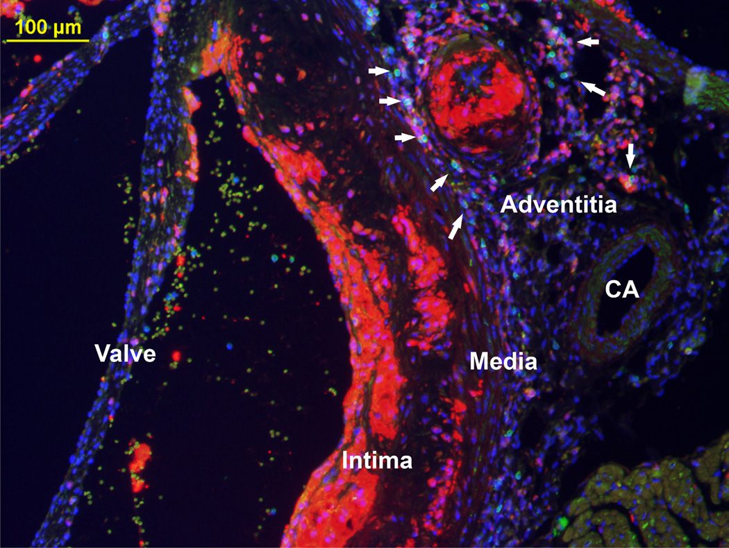
Figure 2. Fluorescent microscopy (10X) of a cross-section from the aortic root of a 24 week old Ldlr−/− mouse fed Western diet for 16 weeks.
Macrophages appear red (anti-Mac-2), T lymphocytes appear green (anti-CD3), and nuclei appear blue (DAPI). The overlay RGB image clearly demonstrates the intima, media, and adventitia with the presence of an atherosclerotic plaque containing macrophages and T lymphocytes. Adjacent to the coronary artery (CA) is an area with T lymphocytes as well as an area of adventitial macrophages.

